New Publication in Bioactive Materials “Studies of Nanoparticle Delivery with In Vitro Bio-engineered Microtissues”
Recently, our group's Ph.D. student, Jinhyung Lee, published a review paper in collaboration with Dr. Yupeng Chen and Dr. Kazunori Hoshino and Mingze Sun titled "Studies of Nanoparticle Delivery with In Vitro Bio-engineered Microtissues" in Bioactive Materials
You can check out the publication here in ScienceDirect
We have summarized the articles below, but more information can be obtained by reading the publication posted above!
THE review focuses on the uses of various types of nanoparticle vehicles such as polystyrene beads, gold nanoparticles, lipid nanoparticles, quantum dots, and others in the application of drug delivery. Nanoparticles that come in all shapes and sizes have been utilized and applied in many areas such as cancer treatment, atherosclerosis treatment, osteoarthritis treatment, neuro-degenerative treatment, and many more. As the nanoparticles are becoming more complex, the efficacy can be tested with the utilization of in vitro bio-engineered microtissues, which is explained more in-depth in the paper.
The paper also discussed ways to synthesize these nanoparticles and modifications used to enhance the efficiency of drug delivery such as the presence of poly(ethylene glycol) (PEG) improving the hydrophilicity of copolymers, reducing the concentration of lactic acid produced after degradation. These modifications or functionalization of the surfaces of the nanoparticles are especially useful because they improve penetration as well as an increase in targeting delivery. With improve penetration, these functionalized particles could change the mechanism of membrane transport and even promote the penetration of drugs to biofilm and biological barriers in vivo.
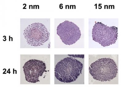
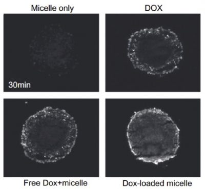
Because there are different types of nanoparticles developed, the easiest way to study the impact of the delivery of these nanoparticles quantitatively is by using engineered tissue models. These models can be bioengineered 3D cell culture models like multicellular spheroids, that can provide a better perspective than conventional monolayer 2D cell culture method, as seen on figure 2 in the paper that showed the penetration of doxorubicin (DOX) to multicellular spheroids and the uptake into the core can be observed within 30 minutes. Figure 3 in the paper shows the penetration of different sized gold nanoparticles with spheroids after incubation of 3 and 24 hours. Figures 2 and 3 can be seen below.

Figure 2 and Figure 3. The penetration of gold nanoparticles into tissues as well as doxorubicin (DOX) into spheroids tissues.
The paper concluded that the current most commonly used methods for the study of nanoparticles are the utilization of multi-cellular spheroids because it can be easily prepared. However, the challenges scientists face currently is that these spheroids syntheses are dependant on the biological characteristics of cells used, therefore, quite unpredictable, in preparing a standard size/ shape/ properties across the board for more uniform testing. Other challenges faced by scientists are the lack of data in the correlation of mechanical characteristics and transport properties, such as the measurement of the elastic modulus allowing simple and direct means for quantitative tissue analysis.
Categories: Uncategorized
Published: August 13, 2020
